Ultrasound
Ultrasound
What Is Diagnostic Ultrasound?
Ultrasound is a non-invasive imaging technique that uses high-frequency sound waves to create real-time images of structures inside the body. At Katranji Hand Center, diagnostic ultrasound is an essential tool in evaluating conditions affecting the hand, wrist, elbow, and upper extremities.
Unlike X-rays, which show bones, ultrasound allows us to visualize soft tissues like tendons, nerves, muscles, and blood vessels—helping guide accurate diagnoses and targeted treatments.
What Ultrasound Can Detect
Ultrasound is especially useful for identifying:
Tendon tears or inflammation (e.g., De Quervain’s tenosynovitis, trigger finger)
Nerve compression or abnormalities (e.g., Carpal Tunnel Syndrome)
Joint effusions (fluid buildup)
Ganglion cysts or masses
Muscle or ligament injuries
Foreign bodies embedded in soft tissue
Dynamic movement issues (seen in real-time with active motion)
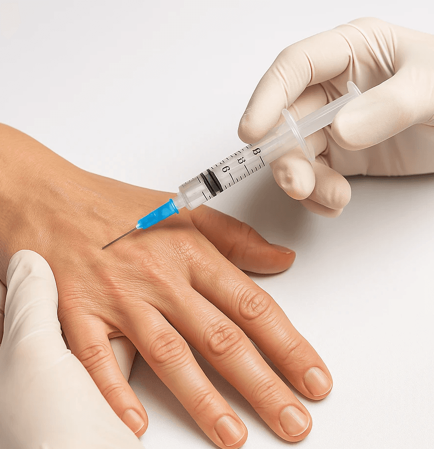
Ultrasound can also guide procedures, such as cortisone injections, ensuring precision and reducing the risk of complications.
Benefits of In-House Ultrasound
At Katranji Hand Center, offering in-house ultrasound provides several advantages:
Immediate imaging during your visit
Real-time feedback for quicker diagnosis and treatment planning
No radiation exposure
Cost-effective and comfortable compared to MRI or CT
Ideal for dynamic evaluation, allowing movement to be assessed during the scan
When Ultrasound Is Recommended
Your provider may recommend an ultrasound if you are experiencing:
Unexplained pain, swelling, or stiffness
Numbness or tingling suggesting nerve entrapment
Lumps or bumps under the skin
Injuries that haven’t healed or continue to cause discomfort
Need for guided injection or aspiration
What to Expect During Your Ultrasound
Painless and quick—typically 10–20 minutes
A small probe (transducer) and gel are used on the skin
You may be asked to move or flex your fingers, wrist, or arm
Results are interpreted directly by your specialist during the same visit
Follow-Up and Treatment
Depending on the results, next steps may include:
Physical or occupational therapy
Splinting or bracing
Corticosteroid injections
Surgical consultation, if structural damage is found
Continued monitoring with follow-up imaging
Ultrasound allows us to personalize your care with precision—getting you back to your daily activities with the most effective treatment plan possible.